
Definition
From the above description of the word, it can be clear that Anisocytosis refers to unequal size of cells. In medicine, the word is used to describe a condition in which the red blood cells (RBC) are of unequal sizes.
Pronunciation
- An-eye-so-cy-toe-sis [1]
Meaning
- Origin- greek [2]
- An- not, no, negative [2]
- Iso- same [2]
- Cyto- cell [3]
- Sis- state or condition [4]
The degree of variation is measured in lab by Red Cell Distribution Width (RDW). If the RDW is high, that means the difference in size of RBCs is more [5]. It is important to remember ‘Width’ in the term RDW does not mean the width or diameter of the red blood cell but the width of the volume curve [6].
See : Red Blood Cell Indices
The reference values for RDW is as follows
-
RDW-SD- 39-46 fL [7] (SD refers to standard deviation in femtolitres)
-
RDW-CV- 11.6- 14.6 % in adult [8] (CV refers to coefficient of variation)
Usually anisocytosis is seen in various anaemias. To correctly diagnose the cause of anaemia we use RDW in conjunction with MCV (Mean Corpuscular Volume). MCV is not the volume of a single RBC, but is the average volume of RBC in the given sample of blood. The reference value of MCV is 80-96 fL/RBC [8].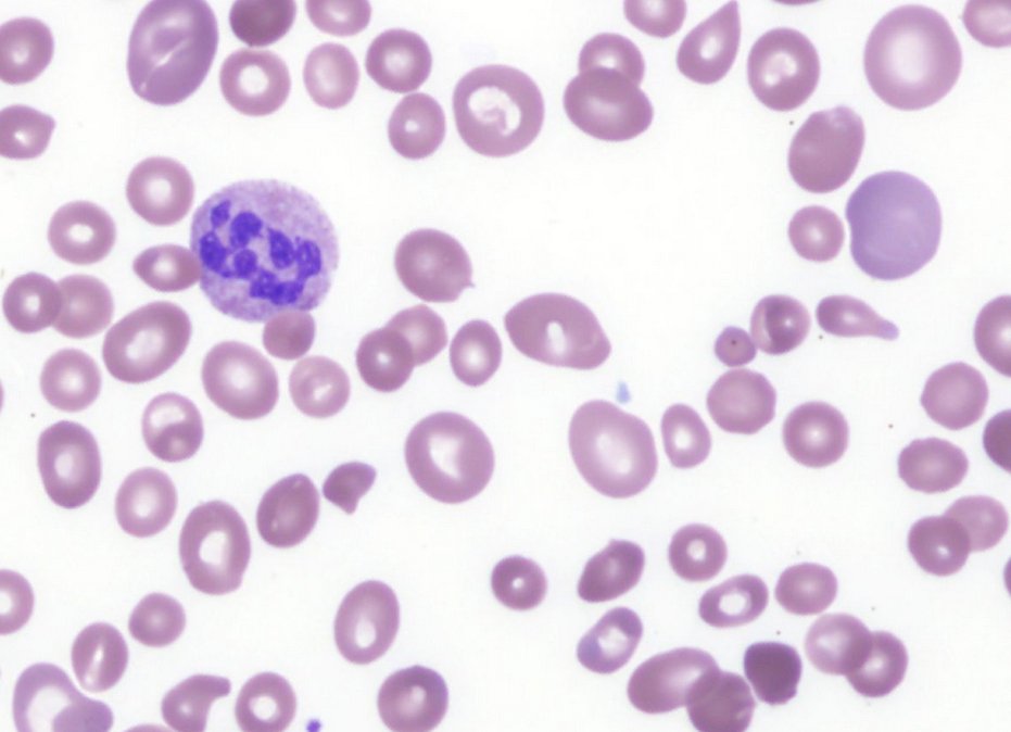
Smear image 1: Peripheral blood film of a patient, with severe Vitamin B12 deficiency, showing marked anisocytosis. Different sizes of RBCs can be noted in the picture.
picture source : bloodjournal.org
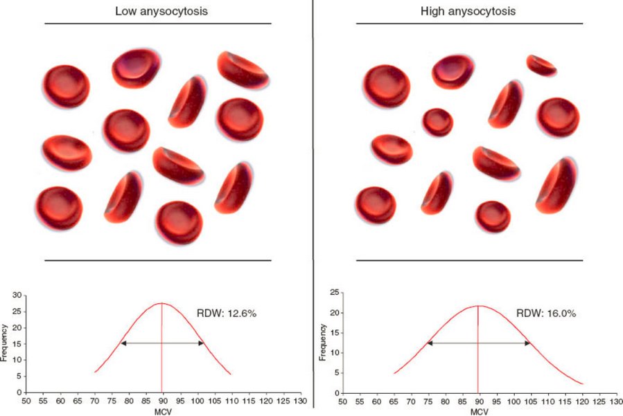
Picture 2 : The variation in low and high anisocytosis can be seen in this image.
Image 2 source : https://www.degruyter.com
Normal RBC
A normal RBC is disc shaped with both of its surface concave (biconcave) and without a nucleus. On a peripheral smear (a glass slide with blood smeared on it for examination under microscope) RBC has a central area that is slightly pale compared to the surrounding area.
This area is approximately one third of the total area of the RBC [9]. The diameter is approximately 6-8 microns [6, 10].
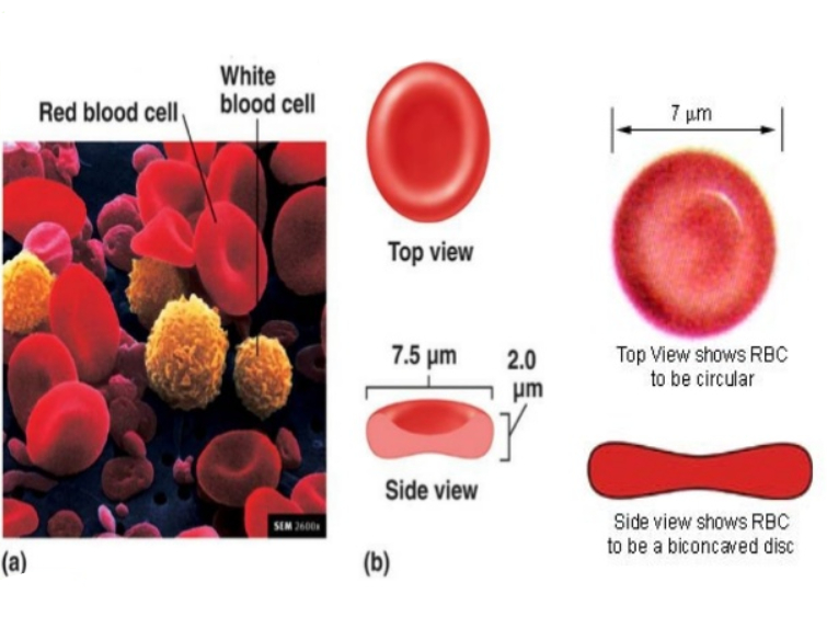
Image showing RBCs and WBCs in the peripheral blood. (b) Image showing the diameter and thickness of RBC.
Types & Causes
On the basis of RDW and MCV measurements, anisocytosis can be
Microcytic
The cell size is decreased than the normal.
- This may be seen in Iron deficiency anaemia, thalassemia, sideroblastic anaemia, lead poisoning, pyridoxine deficiency and sickle cell anaemia[9, 11, 12].
Macrocytic
The cell size is increased than the normal.
- Vitamin B12 deficiency (Megaloblastic anaemia), autoimmune haemolytic anaemia, chronic liver disease, myelodysplastic syndrome, chemotherapy, hypothyroidism, post splenectomy etc.
Normocytic
The cell size is within the normal range.
- Early iron deficiency, early vitamin B12 deficiency, early folic acid deficiency, chronic liver disease, sickle cell disease, myelodysplastic syndrome, dimorphic anaemia.
When the RBCs are of different sizes, the oxygen carrying capacity of these cells varies. This in turn affects the oxygen supply to different tissues [13, 14].
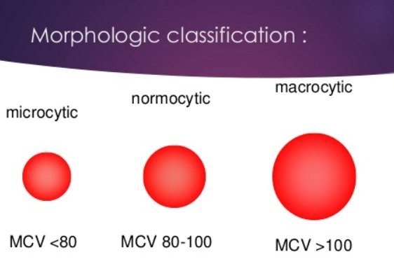
Image 4: Difference between microcytic, normocytic and macrocytic RBCs.
Anisocytosis in Pregnancy
One of the common observations during pregnancy is anaemia, mostly iron deficiency anaemia or folic acid deficiency anaemia. These conditions may present as mild anisocytosis, especially in the early stages of anaemia[15].
Symptoms
The symptoms of anisocytosis are majorly due to the inefficient oxygen carrying capacity of RBCs which result in decreased oxygen supply to the tissues and organs. Pallor of skin, weakness, fatigue, breathlessness and increased heart rate can be seen in individuals with anisocytosis[16, 17, 18, 19].

Image 5: An examination followed by investigations is important when patients presents with symptoms of anaemia.
Diagnosis
The diagnosis of anisocytosis can be made by examining the blood. RDW and MCV estimation is the main stay to a correct diagnosis. Further investigations may be required to find the underlying cause of anisocytosis.
Remember, anisocytosis by itself is not a disease, but an expression of an underlying abnormal condition[22, 23].
Grading of anisocytosis is done based on the degree of variation from the normal size. The grades given are + (Mild/ 1+), ++ (Moderate/2+) or +++ (Severe/3+).
Based on RDW, the grades are
-
+/ Mild/ 1+ Anisocytosis- When RDW= 16 to 18
-
++/ Moderate/ 2+ Anisocytosis- When RDW = 18 to 22
-
+++/ Severe/ 3+ Anisocytosis- When RDW >22
Treatment
Treating anisocytosis can be done only by correcting the underlying cause. If it is due to anaemia, the correct cause of it needs be diagnosed first. Nutritional supplementation with diet modification may help in anaemia due to deficiencies.
But in case of thalassemia a blood transfusion might be what is needed, whereas myelodysplastic syndrome may require a bone marrow transplant[24, 25, 26, 27, 28, 29].
Reference:
- https://www.merriam-webster.com/medical/anisocytosis
- http://wordinfo.info/unit/2389/ip:10
- https://medical-dictionary.thefreedictionary.com/cyto-
- https://medical-dictionary.thefreedictionary.com/-sis
- https://emedicine.medscape.com/article/2098635-overview
- https://en.wikipedia.org/wiki/Red_blood_cell_distribution_width
- Briggs C, Bain BJ. Basic Haematological Techniques. Bain BJ, Bates I, Laffan M, Lewis SM. Dacie and Lewis Practical Haematology. 11th ed. Philadelphia, PA: Churchill Livingstone/Elsevier; 2012. chap 3.
- Vajpayee N, Graham SS, Bem S. Basic Examination of Blood and Bone Marrow. McPherson RA, Pincus MR. Henry’s Clinical Diagnosis and Management by Laboratory Methods. 22nd. Elsevier/Saunders: Philadelphia, PA; 2011. 30.
- http://laboratoryinfo.com/variations-in-red-blood-cell-morphology/
- https://www.labce.com/spg469710_normal_red_blood_cell_rbc_morphology.aspx
- https://en.wikipedia.org/wiki/Anisocytosis
- https://emedicine.medscape.com/article/2098635-overview#a2
- https://www.urmc.rochester.edu/Encyclopedia/Content.aspx?ContentTypeID=160&ContentID=34
- http://biology.about.com/od/humananatomybiology/ss/red-blood-cells.htm
- Friel LA. (n.d.). Anemia during pregnancy. http://www.merckmanuals.com/home/women-s-health-issues/pregnancy-complicated-by-disease/anemia-during-pregnancy
- http://www.nhs.uk/conditions/Anaemia-iron-deficiency-/Pages/Introduction.aspx
- https://www.nhlbi.nih.gov/health/health-topics/topics/ida/signs
- http://www.webmd.boots.com/a-to-z-guides/polycythaemia
- https://www.healthline.com/health/anisocytosis#overview1
- https://www.healthline.com/health/anisocytosis#causes3
- https://medinfo.org/anisocytosis-symptoms-causes-diagnosis-treatments.html
- http://www.bloodmed.com/home/ebook.asp?book=0865427178&id=2
- http://www.hkimls.org/qapeduhsm-rbc-mor.htm
- https://www.healthline.com/health/anisocytosis#treatment5
- https://books.google.com/books?id=M92T5q1hzRoC&dq
- https://en.wikipedia.org/wiki/Iron-deficiency_anemia
- https://en.wikipedia.org/wiki/Red_blood_cell
- https://www.nhlbi.nih.gov/health/health-topics/topics/ha
- https://emedicalhub.com/anisocytosis/